Preimplantation WGS(Whole Genome Sequencing)Testing (PGT-WGS)
AboutThe Preimplantation Whole Genome Sequencing Testing (PGT-WGS) allows for the examination of fertilized eggs for mutations that cause congenital diseases at the single nucleotide level before pregnancy. Furthermore, since fertilized eggs (embryos) can be cryopreserved, you can take your time to carefully consider which embryos to implant without worrying about time constraints.
Approximately 3-5% of newborns are born with congenital diseases (congenital abnormalities). 25% of these cases are due to chromosomal abnormalities (such as trisomy, monosomy, translocation, etc.). Congenital diseases caused by a single gene account for around 20%, which is similar to the percentage caused by chromosomal abnormalities. Furthermore, congenital abnormalities caused by multiple factors (a combination of environmental and genetic mutations) reach up to 50%. Therefore, the proportion of congenital diseases caused by genetic mutations may be higher than those caused by chromosomal abnormalities.
Moreover, chromosomal abnormalities (such as trisomy or monosomy) become more frequent as age increases. Therefore, in younger individuals, there are reports suggesting that congenital diseases caused by genetic mutations may pose a higher risk compared to chromosomal abnormalities.
This Preimplantation Whole Genome Sequencing (PGT-WGS) test examines all genes at the level of base sequence in fertilized eggs before pregnancy, allowing the detection of mutations that may cause diseases, including newly occurring mutations, not only from parental inheritance.Based on the results of this test, it is possible to select embryos for pregnancy and perform thawed embryo transfer by returning cryopreserved embryos to the uterus.
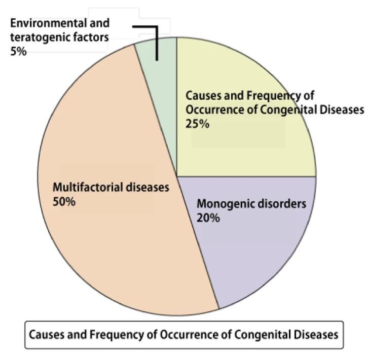
Congenital disorders include more than just trisomy and monosomy.
It is known that 3-5% of babies are born with congenital diseases.
25% of these are caused by chromosomal abnormalities (trisomy, monosomy, translocation). Trisomy is a condition in which a pair of chromosomes becomes three. Human chromosomes usually have a pair of two chromosomes. However, in trisomy, an extra chromosome is present. A well-known congenital disease with trisomy is Down syndrome. In Down syndrome, there are three copies of chromosome 21. Monosomy is the opposite, in which there is only one chromosome missing. For example, in Turner syndrome, there is monosomy with only one X chromosome, which is a sex chromosome.
Approximately half of congenital diseases are caused by multifactorial diseases (a combination of genetic mutations and environmental factors). As a result of advances in genome analysis, mutations related to diseases have been found, even in diseases that were not previously thought to be caused by genetic mutations, such as lifestyle-related diseases. For this reason, many diseases are now considered to be multifactorial (complex) genetic disorders in which individual gene mutations alone do not lead to disease, but develop when multiple mutations are combined.
On the other hand, the percentage of congenital diseases caused by specific gene mutations is 20%, which is slightly lower than chromosomal abnormalities such as trisomy. However, considering that multifactorial diseases are influenced by gene mutations, congenital diseases caused by some kind of gene mutation may occur much more frequently than chromosomal abnormalities.
In younger people, genetic disorders caused by single gene mutations are more common
It is known that the frequency of numerical chromosomal abnormalities depends on the woman's age. As a woman gets older, the percentage of eggs with numerical chromosomal abnormalities increases. Therefore, as a woman gets older, the possibility of fertilized eggs and fetuses having trisomy or monosomy increases. Therefore, NIPT (new type prenatal diagnosis), chorionic villus sampling, and amniocentesis are recommended for women over 35 years old.
The risk of numerical chromosomal abnormalities (trisomy and monosomy) is not as high for younger people as it is for older people. Rather, there is research that shows that congenital diseases caused by single gene mutations are more frequent than trisomy and monosomy for younger people.
Diseases caused by single gene mutations rarely cause abnormalities in the morphology of the fetus, and abnormalities may be found by ultrasound examination. However, regular PGT-A (PGS), NIPT, chorionic villus sampling, and amniocentesis cannot detect single gene mutations. As a result, abnormalities are almost always found after birth.
Carriers of genetic disorders
Diseases caused by a single gene mutation are inherited in a recessive manner. In this inheritance pattern, if a normal gene and a gene with a disease-causing mutation are paired, the disease will not develop. This state is called a carrier. The disease will develop only when both genes with the disease-causing mutation are present.
It is not possible to determine whether a person is a carrier from appearance, as the person will not develop the disease. Therefore, if two carriers of the same genetic disease happen to become a couple, there is a 25% chance that they will inherit the gene with the disease-causing mutation from their parents and develop the disease.
If you have been screened for carriers in advance and know that you are a carrier, you can use PGT-M (preimplantation genetic testing) to select an unaffected embryo and become pregnant.
Mutation frequency
However, carrier screening is not a panacea. Genetic mutations that cause diseases are found every day, so carrier screening may not cover all genetic mutations that cause diseases. Furthermore, mutations may occur, and even if both parents have normal genes, children may have congenital abnormalities.
The probability of a mutation occurring is very small, at1×10-8per base. However, the human genome consists of3×109bases. Therefore, new mutations are calculated to occur in approximately 30 bases (30 locations) per generation. Furthermore, research has shown that the probability of mutations in men increases with age, just like chromosomal abnormalities in eggs.
Not all of these will cause congenital diseases, but depending on the location of the mutation, there is a possibility that congenital diseases may develop. Naturally, these new mutations cannot be prevented by carrier screening or PGT-M.
Methods
MethodsPGT-WGS Testing Method
In PGT-WGS, fertilized eggs are first created through in vitro fertilization (IVF). After about five days of culturing in vitro, the fertilized eggs undergo cell division and differentiate into cells that will become the future placenta and cells that will become the fetus. A portion of the cells destined to become the placenta is then sampled and used for PGT-WGS testing. Since it takes time to obtain the results of PGT-WGS, the fertilized eggs (embryos) are cryopreserved until the test results are available.
DNA is extracted from the sampled cells. This extracted DNA is then subjected to next-generation sequencing (NGS) to examine the entire genome's base sequence at a single-base level. The read base sequences are compared with the parents' base sequences to check for mutations that may cause genetic diseases.
The genome contains exons, which are the blueprints for proteins, and non-coding regions such as introns that connect them and do not serve as blueprints for proteins. Mutations in the exons, which are blueprints for proteins, often result in the inability to produce normal proteins. Although non-coding regions do not serve as blueprints for proteins, they include parts that control the amount, location, and timing of protein production. Therefore, mutations in non-coding regions can also lead to genetic diseases. This WGS examines the entire genome's base sequence, including non-coding regions, for any mutations.
Once the results of PGT-WGS are available the desired embryo for pregnancy is selected based on the test results, and frozen embryo transfer (FET) is performed to return the cryopreserved embryo to the uterus. Embryos can be cryopreserved almost indefinitely. Therefore, unlike prenatal tests such as NIPT, chorionic villus sampling, and amniocentesis, embryos can be considered for transfer without worrying about time constraints, allowing for satisfactory decision-making.
Limitations of PGT-WGS
However, PGT-WGS also has its limitations. Some mutations have unclear clinical impacts, and future research may change the interpretation of these mutations. Thus, mutations currently considered unrelated to diseases may later be found to be associated with diseases, or vice versa. Additionally, in cases of mosaicism, where only some cells in the body have mutations, there is a risk of missing these mutations, or mutations that cause diseases may be detected without the disease actually manifesting. Furthermore, genetic diseases vary in penetrance and expressivity depending on the mutation, meaning that a detected mutation may not always lead to disease manifestation. NGS is not yet a perfected technology. While the accuracy of reading base sequences is very high, it is not 100%, so there is a possibility of missing mutations or incorrectly identifying potential disease-causing mutations.
Costs
Fee DetailsThe total cost consists of the basic fee, the testing fee for embryos, and the WGS data storage fee.
- Basic fee (includes WGS testing for the couple and one year of WGS data storage): ¥300,000 (¥330,000 incl. tax)
- Embryo testing fee (per embryo): ¥250,000 (¥275,000 incl. tax)
- WGS data storage fee (per year) Couple’s WGS data: ¥23,000 (¥25,300 incl. tax)
Embryo WGS data (per sample): ¥14,000 (¥15,400 incl. tax)